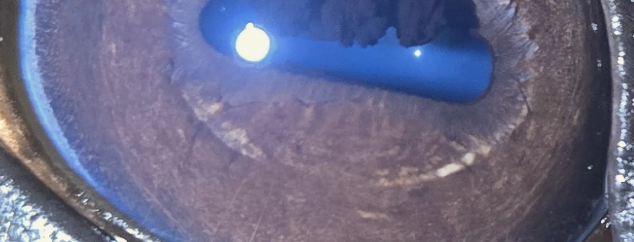
What it does
- Indicates inherited constitutional strengths and weaknesses
- Shows areas of tissue under stress or congestion
- Flags accumulation the body has not eliminated
- Records old injury and scarring
- Suggests where to investigate next
The Method
This is the sequence Ellen Collinson built by correlating iris markings against what veterinary surgeons actually found. It is what the diploma teaches, and it is what happens behind every £70 report.
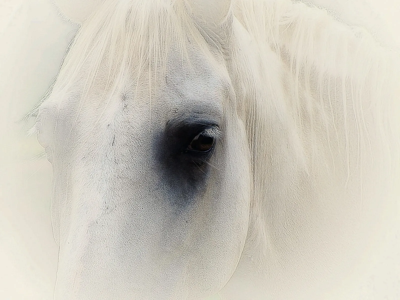
Nothing touches the horse. No sedation, no restraint beyond a headcollar, no blood, no stress. A single clear image of each eye is the entire physical component of an iris analysis, which is precisely why it can be done on horses that will not tolerate anything else.
The iris is read as a clock face. Every sector corresponds to a region of the body, and the two eyes report differently. The first task is not interpretation but orientation: establishing exactly where on the map a marking sits.
How tightly or loosely the fibres sit tells you about the integrity of the tissue underneath. Colour and pigment tell you about accumulation. Read them in that order. Structure is constitutional, whereas colour is often acquired and therefore changeable.
A marking means little without the animal in front of you. Findings are checked against history, diet, workload and presenting signs. This is where a competent practitioner separates from someone reading patterns off a chart.
A report that only describes is worthless. The output is a plan: herbal support matched to indication, feeding adjustments, management changes, and where warranted, a clear instruction to call the vet.
Reading the eye
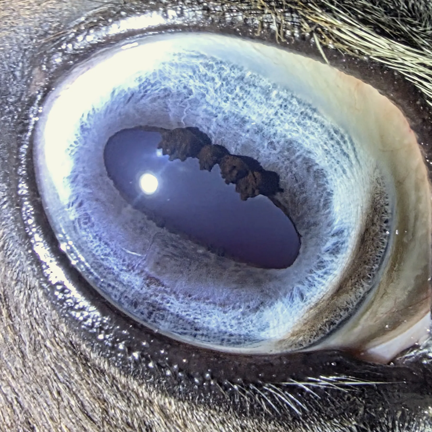
The density and regularity of the fibres running out from the pupil. Hereditary traits and behavioural patterns are clearly marked within the fibre patterns.
The band immediately around the pupil. Its regularity carries a great deal of information about the digestive tract.
The dark scalloped bodies at the pupil margin. Distinctive to horses, and one of the first structures you learn to read properly rather than mistake for a marking.
Discolouration and deposits indicate accumulation: what the body has not managed to eliminate, and where it has chosen to store it instead.
Offers insight into congestion, blockages and mineral imbalances.
This is an illustration, not a chart. Reading the full map, both eyes, and orienting yourself in any photograph is built up across the whole diploma.
Scope of practice
We are deliberate about this. A practitioner who oversells iridology damages every other practitioner, and eventually the horse.
Equine iridology is a complementary discipline and is not statutorily regulated in the UK. We say so plainly, and we teach every student to work alongside veterinary surgeons rather than in place of them.
Where it came from
Iridology in humans has been studied for centuries. Its arrival in the equine world is more recent and considerably stranger.
Syd Mercer, an English practitioner trained by a veterinary surgeon during the First World War, used equine eye diagnosis on racehorses, reportedly with enough success to interest people who cared a great deal about the outcome of races.
Ellen Collinson brought rigour to it. She had already studied human iridology when she turned to horses, and she did the unglamorous work: recording markings, then attending post-mortems to see whether the eye had been right.
Next
Ten modules if you want to practise it yourself. A single report if you just want an answer.
£1,500 for the diploma, or five monthly payments of £300. £70 for a single report.
Not sure which? and she'll talk it through with you.